
Figure 3.
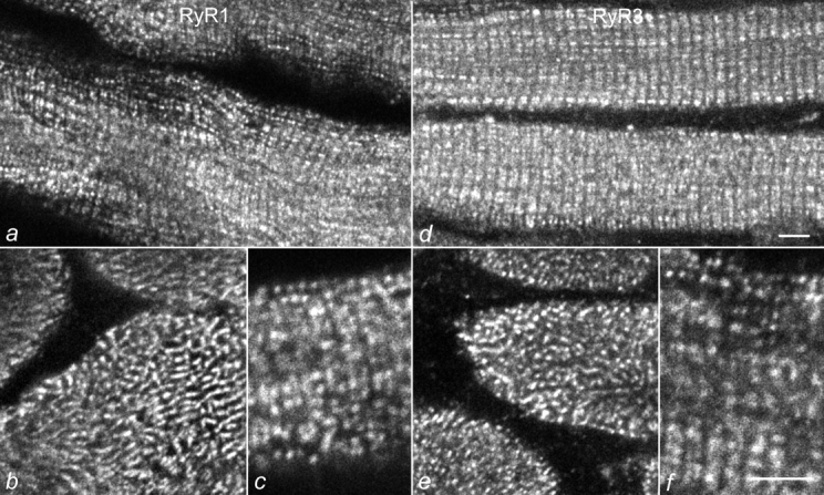
Colocalization of RyR1 and RyR3 in young mouse skeletal muscle. Sections of D15 normal mouse diaphragm muscles were immunolabeled with specific antibodies against RyR1 (a–c) and RyR3 (d–f). Both antibodies show the typical labeling pattern of triad proteins: double rows of fluorescent dots representing the pairs of triads on either sides of the Z-line or the meshwork between the myofibrillar bundles, in longitudinally (a, c, d, and f) and cross-sectioned (b and e) muscle fibers, respectively. Bars, 5 mm.