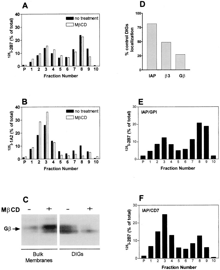
Figure 5.
Effect of cholesterol depletion on DIGs localization of signaling components. A–D, OV10 cells expressing IAP treated with or without 10 mM MβCD before Brij lysis and sucrose density centrifugation. Distribution of 125I-labeled anti-IAP mAb 2B7 (A) or anti-β3 mAb 1A2 (B) was determined as described in Fig. 4. C, Bulk membranes (fractions 2–4 combined from a sucrose gradient) and DIGs containing fractions (fractions 7–9 combined) were analyzed by SDS-PAGE and Western blotting using rabbit antibodies specific for Gβ after sucrose density fractionation of unlabeled cells. D, The percent of IAP, β3, and Gβ remaining in the DIGs-containing fractions 7–9 after MβCD treatment. Percentage of total counts was used to determine the amount of IAP or β3 present, and densitometry of Western blots was used to determine the amount of Gβ present. E and F, Distribution of IAP Ig domain-expressing molecules after sucrose density centrifugation of Brij-lysed OV10 cells expressing IAP/GPI (E) or IAP/CD7 (F) was determined as described in Fig. 4.

