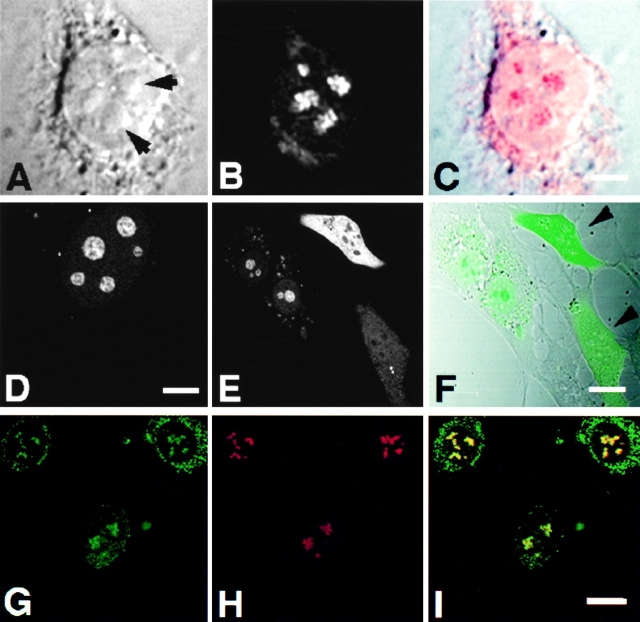
Figure 9.
Subnucleolar localization patterns of Rpp14. 3T3 fibroblasts were subjected to indirect immunofluorescent analysis using anti-Rpp14 antibodies (A–C). C is an overlay of A and B. Fibroblasts transfected for 48 h with pEGFP-Rpp14 were examined under confocal microscope (D–F) before fixation and immunofluorescence analysis with anti-Rpp29 antibodies (G–I). F is an overlay of DIC (not shown) and E; two cells with high fluorescent signal are indicated by arrowheads. I is an overlay of G and H. Intense yellow color is seen in nucleoli. Bars: C and D, 3.3 μm; F and I, 10 μm.

