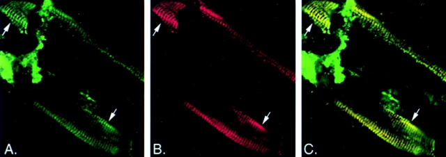
Figure 2.
Confocal images of a wild-type adult worm fragment stained for intermediate filaments (green) and myotactin (red). (A) Intermediate filaments are concentrated in thin bands running circumferentially around the worm (arrows). The intermediate filament-dependent staining within each band is not uniform but is associated with discrete punctate structures within the band. (B) Like intermediate filament-dependent staining, myotactin-dependent staining is associated with punctate structures that are organized into bands (arrows). (C) Merging of the images shown in A and B shows the punctate patterns seen with the two antibodies are coincident and thus produce the yellow fluorescence.

