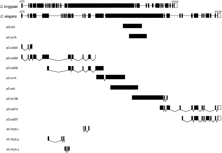
Figure 3.
Schematic diagram of the partial myotactin cDNAs and genomic clones. The top two lines represent C. briggsae and C. elegans genomic clones, respectively. Exons are depicted as filled boxes and introns as lines. pG-cc9 is the genomic clone isolated using the MH46 antibody as a probe (see Materials and Methods) and pC-ccXX(X) are the overlapping cDNA clones. RT-PCR-1, 2 and 3 (see Materials and Methods) were sequenced to confirm the intron–exon boundaries between exons 5 and 6, 6 and 7, 7 and 8, 8 and 9, 9 and 10, 12 and 13, and 13 and 14. The open boxes at the 3′ ends of pC-cc214, pC-cc307, and the C. elegans and C. briggsae genes represent noncoding sequence. The positions of the predicted translational start (ATG) and the stop codon (TAA) are indicated. pC-cc503 contains eight bases at the 5′ end identical to the eight 3′ bases of the SL1 spliced leader sequence. Note that exons 6–9, 13, 19A, 19B, 24 and 25 are all differentially spliced. The combinations with which these exons are used has not been determined. The complete sequence of the myotactin cDNAs is attainable through Genbank. Two cDNA sequences were submitted: Form A includes exon 19A but excludes exons 19B, 24, and 25 (accession number AF148954) and form B includes exons 19B, 24, and 25 but excluded 19A (accession number AF148953). The two forms reflect the differences observed between cDNA pC-cc214 and pC-cc307.

