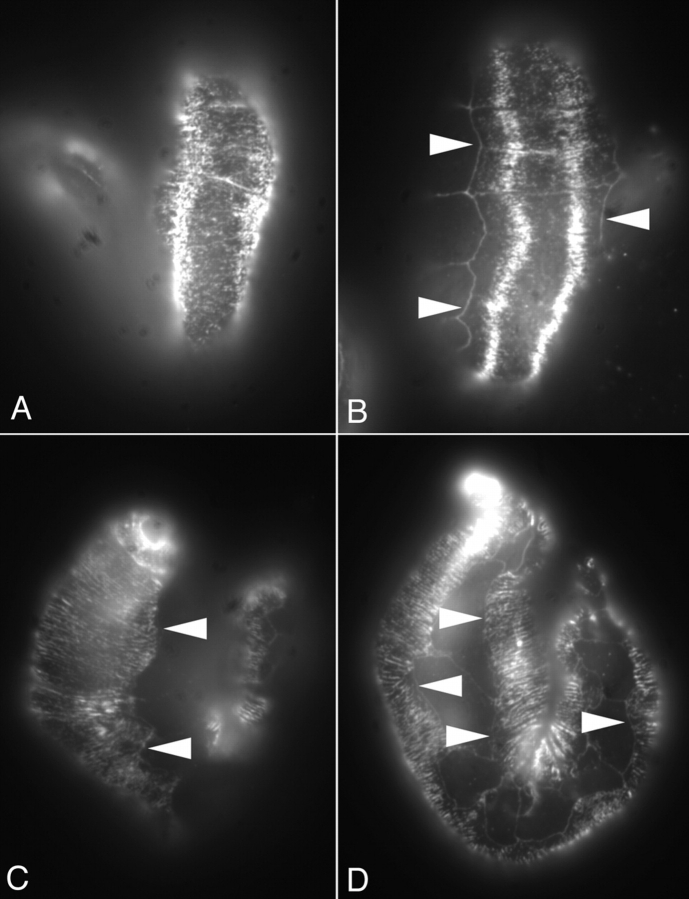
Figure 8.
Wild-type and myotactin mutant embryos stained for intermediate filaments. Mixed stage embryos were fixed for immunofluorescence and labeled with MH4 (intermediate filament protein) and MH27. MH27 marks the boundaries between hypodermal cells and appears as a grid pattern on the embryos in B and D. (A and B) Dorsal view of a wild-type (A) and a st456 homozygous (B) embryo. In both embryos, the MH4-dependent signal is concentrated in the regions of the hypodermis adjacent to muscle. (C) Dorsal view of a st456 homozygote older than the one shown in B. MH4-dependent fluorescence is seen all through the dorsal hypodermis. (D) Lateral view of an embryo at a similar stage to the one shown in C. The MH4-dependent staining extends to the boundaries between the dorsal or ventral hypodermis and the seam hypodermis. The fluorescence pattern seen in C and D is one of circumferentially oriented bands. Arrowheads mark the boundaries between dorsal or ventral and seam hypodermis.

