
Figure 7.

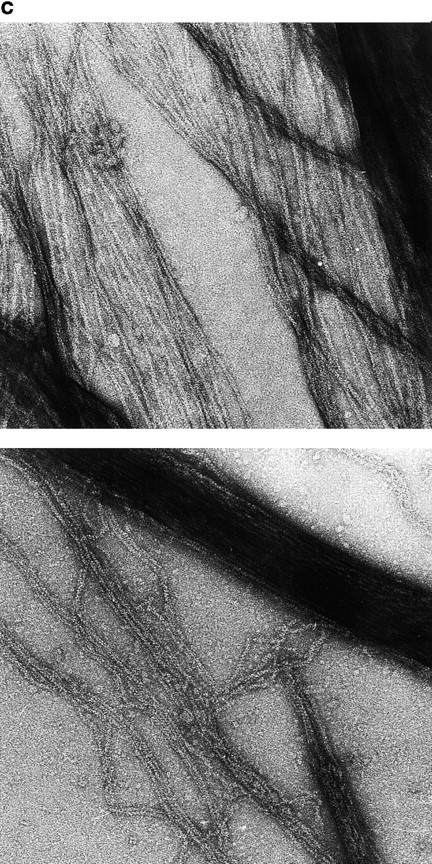
VASP induces polymerization of G-actin into F-actin bundles. (a) Pyrenyl-actin fluorescence measurements. 1 μM pyrenyl-labeled MgATP-G-actin was supplemented, at time indicated by the vertical arrow, with eukaryotic recombinant VASP at the following concentrations (μM): a, 0; b, 0.25; c, 0.5; d, 1; e, 2; f, 1 μM VASP + 0.1 M KCl; g, 0 VASP, 2 μM GST-EVH2. The fluorescence of 1 μM pyrenyl-G-actin is equal to 1 by convention. The horizontal arrow indicates the fluorescence measured for 1 μM F-actin polymerized in standard F buffer (0.1 M KCl, 1 mM MgCl2 added to G buffer). (Inset) Extent of fluorescence change at the end of the polymerization process of 0.7 μM MgATP-G-actin induced by VASP at the indicated concentrations. Fluorescence was measured 1 h after the preparation of the samples. Final conditions were: 5 mM Tris-Cl−, pH 7.5, 0.2 mM ATP, 0.1 mM CaCl2, 1 mM DTT, 0.2 mM EGTA, 50 μM MgCl2, 2 mM Hepes, 15 mM NaCl, 1% glycerol, 20°C. (b) Turbidity measurements at 310 nm. 1 μM MgATP-G-actin (same actin solution as in a) was supplemented, at time 0, with eukaryotic recombinant VASP at the following concentrations (μM): a, 0; b, 0.29; c, 0.58; d, 1.16; e, 1.74; f, 2.34; g, 0 VASP, 2 μM GST-EVH2; h, 1.16 μM VASP + 2 μM GST-EVH2; i, 1.16 μM VASP + 0.05 M KCl; j, 1.16 μM VASP + 0.1 M KCl; k, no actin, 1.16 μM VASP. Ionic conditions were as in a. Temperature was 20°C. Optical path length = 1 cm. (c) Actin bundles assembled from Mg-actin in the presence of VASP. Samples of MgATP-G-actin (1 μM) preincubated for 10 min with 1 μM VASP were negatively stained and observed in the electron microscope. (Top) Bacterial recombinant VASP. (Bottom) Eukaryotic recombinant VASP.