
Figure 2.
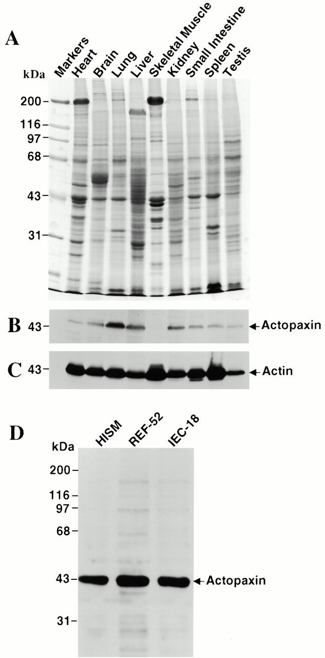
Expression of actopaxin protein in various tissues and cell lines. Organs were dissected from a healthy adult rat, homogenized, and lysed in SDS-PAGE sample buffer. 50 μg of each lysate was loaded per lane. (A) Coomassie blue staining of the gel. (B) Western blotting of an identical gel shown in A probed with the actopaxin antibody. (C) The same blot was stripped and reprobed with an actin antibody to indicate relative loading among lanes. (D) Western blot of total lysates (50 μg) from HISM cells, rat embryo fibroblasts (REF-52), and rat intestinal epithelial cells (IEC-18) probed with the actopaxin antibody.