Figure 1.

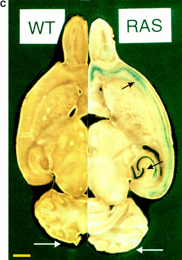
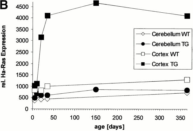
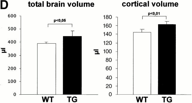
Generation of Ras-transgenic mice: developmental expression of constitutively activated Ras and increased brain volume in adult mice. (A) Schematic diagram of the bicistronic transgene construct. The Ras-TG expression was driven by the neuron-specific promoter of the synapsin I gene. The structural genes of the construct consisted of the gene for constitutively activated V12-Ha-Ras and the lacZ gene. An “internal ribosomal entry site” (IRES) is located downstream of the Ras stop codon. The bicistronic mRNA generated by this construct is terminated at the polyadenylation site of LacZ. Synapsin I promoter was from Rattus norvegicus; V12-Ha-Ras, human Val12–Ha-Ras oncogene (R. Jaggi, Inselspital Bern, Bern, Switzerland); IRES of encephalomyocarditis virus (J.E. Majors, Washington University School of Medicine, St. Louis, MO); LacZ was from Escherichia coli. (B) Developmental Ha-Ras expression in various brain regions and at different ages of Ras-transgenic (synRas-TG) mice and wt littermates. Homogenates of brain cortices or cerebella derived from synRas-TG mice and wt littermates were analyzed by Western blotting using an antibody recognizing endogeneous and activated transgenic Ha-Ras. Ras-TG expression was hardly detectable prenatally. In the cortex, there was a rapid postnatal increase reaching a 4.2-fold expression of activated Ras-TG, relative to endogeneous Ha-Ras at 40 d. Only moderate postnatal Ha-Ras increases were seen in wt cortex or in cerebella of wt or synRas-TG mice. Data are the means of two independent experiments, as obtained from densitometric measurements. (C) Horizontal sections of paired brain hemispheres derived from wt (left) or synRas-TG (right) mice after fixation and X-gal staining. Note the intense expression of the transgene in the region of neuronal cell somata of the hippocampus and also in the cortex (black arrows). The cortical brain volume is enlarged, leading to a caudal displacement of the cerebellum (compare white arrows). Bar, 1.8 mm. (D) Total brain volume and cortical brain volume are enlarged as determined from 13 contiguous coronal slices. Morphometric evaluation of wt (n = 5) or synRas-TG (n = 5) mice was performed by using NMR imaging and a semiautomatic imaging software on a Silicon Graphics Indy computer.


