Figure 5.

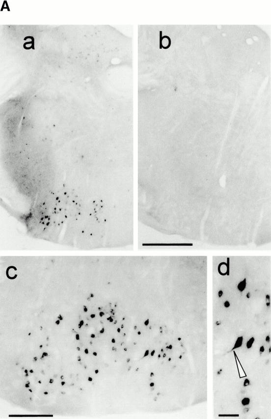
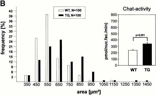
Characterization of Ras-transgenic facial motorneurons. (A) Ras-TG expression in motorneurons of the facial nucleus in adult mice (6 mo old). (a and b) Low-power photomicrographs of coronal sections through the brainstem of a synRas-TG animal (a) and a wt littermate (b). At the level of the genu of the facial nerve, hybridization with lacZ mRNA reveals intense signals in N7 motorneurons and in subsets of neurons in other brainstem structures in the synRas-TG, but not the wt mouse. (c and d) Higher magnification of the facial nucleus at a more posterior level in the synRas-TG animal reveals intensely lacZ mRNA–expressing neurons. The Ras-TG expression varies between the individual neurons, and the reaction product may extend into primary dendrites (arrow head). No signals were observed in glia cells. Bars: (a and b) 200 μm; (c) 50 μm; (d) 20 μm. (B) Size-frequency distribution of facial motorneurons. The average facial motorneuron size is enlarged in synRas-TG mice. Insert: Chat activity in facial nuclei of synRas-TG mice and wt littermates. Expression of Ras-TG protein in facial motorneurons results in a significant increase in Chat activity.

