Figure 6.
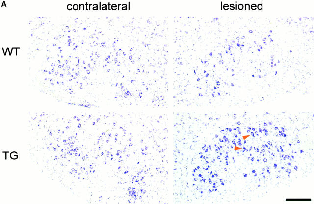
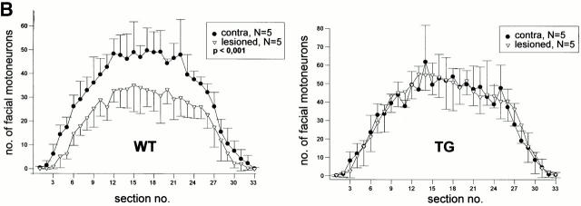
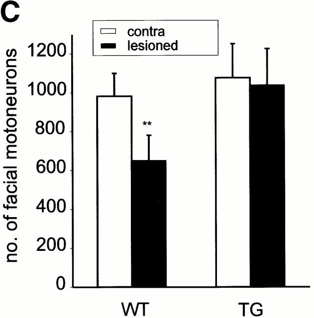
Protection of facial motorneurons from axotomy-induced degeneration. (A) Representative Nissl-stained sections of axotomized mice. The ipsilateral section of the wt mouse shows loss of facial motorneurons compared with the contralateral section, whereas the facial motorneuron number of the synRas-TG mouse remains equal on both sides. Arrowheads indicate well-preserved motorneurons with nucleoli in lesioned synRas-TG mice. Bar, 50 μm. (B) The number of facial motorneurons per section. Facial motorneurons of one out of three sections were counted. The first section is determined by the appearance of the first counted facial motorneuron. For better visualization, the ipsilateral distribution curve was centered within the contralateral distribution curve. Determination of wt facial motorneuron numbers reveals severe axotomy-induced degeneration along the total ipsilateral nucleus length, which cannot be detected in the synRas-TG mice. (C) The number of counted facial motorneurons per nucleus. Facial motorneurons of one out of three sections were counted. Total numbers of facial motorneurons per nucleus were not extrapolated. Analysis of counted facial motorneuron numbers reveals full protection of facial motorneurons from axotomy-induced degeneration by the transgene.


