Figure 7.

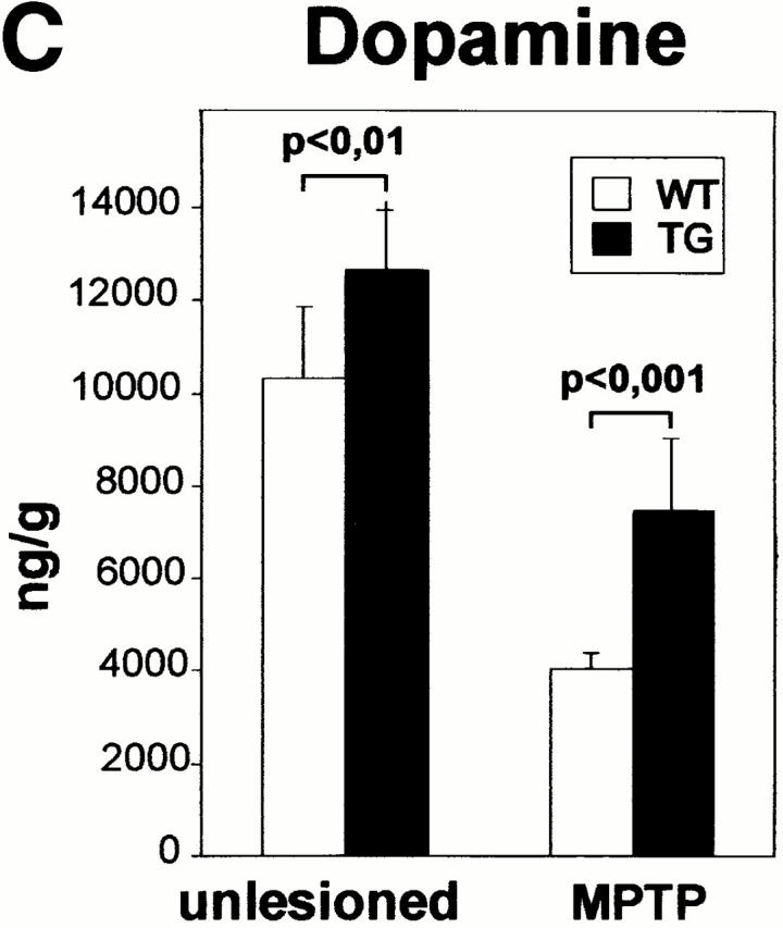
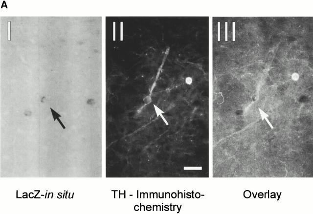
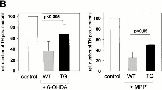
Characterization of transgenic midbrain dopaminergic neurons and protection against neurotoxin-induced degeneration. (A) Expression of the Ras-TG in dopaminergic neurons of the substantia nigra. (I) A coronal section through the midbrain at the level of the substantia nigra was hybridized with a lacZ riboprobe and showed staining in a subset of neurons. (II) Immunohistochemical analysis of the same section using a monoclonal antibody against TH (Roche) and a Texas red–labeled secondary antibody shows TH expression in the neuron expressing the transgene. Bar, 20 μm. (III) Overlay of the immunofluorescence picture and the brightfield micrograph, indicating that the Ras-TG transcript is found in the TH-positive neuron. (B) Effect of 6-OHDA or MPP+ treatment on the number of cultured midbrain dopaminergic neurons derived from synRas-TG mice or their wt siblings. Midbrain dopaminergic neurons were derived from E14 embryos and cultured under serum-free conditions. After treatment with MPP+ (1 μM) for 36 h or 6-OHDA (150 μM) for 90 min, cells were fixed with methanol. Dopaminergic neurons were identified using a monoclonal antibody against TH (Roche), a biotin-labeled secondary antibody (Sigma-Aldrich), and a streptavidine–FITC conjugate. After counting dopaminergic neurons, the genotype was determined by Southern blot analysis. Treatment with the toxin resulted in degeneration of the dopaminergic neurons. This toxin-induced degeneration was markedly reduced in cultures derived from synRas-TG mice. The numbers of nondopaminergic neurons remained unchanged (data not shown). (C) Determination of dopamine after in vivo treatments with MPTP in synRas-TG mice and their wt siblings (see Materials and Methods). In the synRas-TG mice, the MPTP-induced decrease of dopamine contents was profoundly reduced.


