
Figure 3.
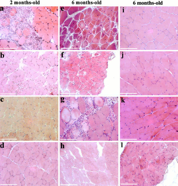
Histologic analysis of capn3-deficient mice. H&E-stained transverse muscle cryosections (7 μm) from mice of 129Sv genetic background. (a–d) 2-mo-old mice; (e–l) 6-mo-old mice. Dystrophic changes are evident in psoas (a and e), soleus (b and f), deltoid (c and g), and tibialis anterior (d and h), whereas quadriceps (i), gastrocnemius (j), and triceps (k) present a normal aspect. Diaphragm in l presents very slight abnormalities. The main features encountered are fibers with internal nuclei (b–f, and h) or area of infiltration of mononuclear cells (a and g). A cluster of small regenerating fibers can be seen in e. Bars, 50 μm.