
Figure 5.
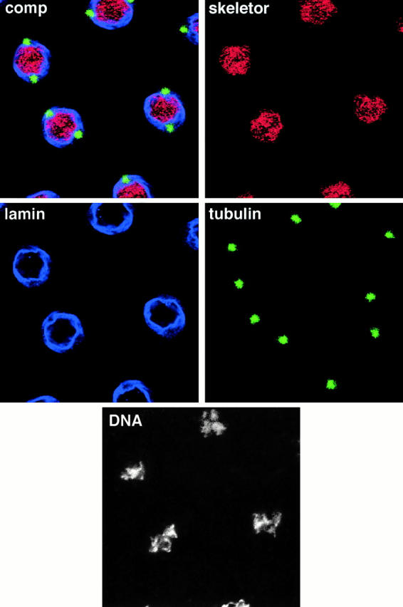
Localization of skeletor, α-tubulin, lamin, and DNA at late prophase in Drosophila embryos. Quadruple labelings of prophase nuclei using mAb1A1 to visualize skeletor (red), anti-lamin antibody to visualize the nuclear lamina (blue), anti–α-tubulin antibody to visualize the microtubules (green), and Hoechst to visualize the DNA (white) reveal that the skeletor spindle has initiated formation before nuclear lamina breakdown and before microtubules can be detected within the nuclear space. The composite panel (comp) shows a merged image of the skeletor, lamin, and tubulin channels, all of which were imaged at the same plane. The DNA panel was imaged from a slightly deeper plane of section, which better represents the condensation state of the DNA.