
Figure 7.
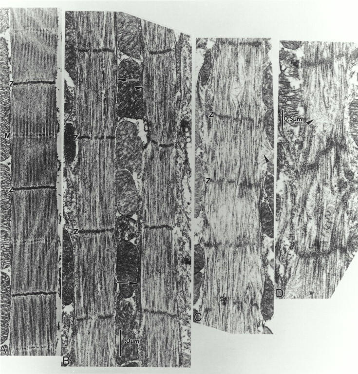
Rapid, progressive sarcomere degeneration in fln0 adult IFM. Electron micrographs of (A) wild-type sarcomeres (B–D) fln0 sarcomeres along the same myofibril in longitudinal 25-nm sections of adult IFM within 12 h after eclosion. In contrast to the relatively well-ordered sarcomeres in pupa (see Fig. 5), adult fln0 sarcomeres are severely disordered. There appears to be a gradual increase in disorder toward one end of a fiber. (B) Even in the best-ordered fln0adult sarcomeres, the M line has disappeared and the Z band tends to fragment. Peripheral bundles of thin filaments (arrowhead) and very long thick filaments (∼10 μm, arrows) are evident. These well-ordered sarcomeres have already shortened to ∼3.1-μm long, about the same length as wild type. (C) Same myofibril as B, but ∼50 μm closer to one end. Note increase in disorder. Z bands are more fragmented and sarcomeres are shorter (∼2.0 μm). In some regions, thick filaments are missing or appear fragmented. Thin filament “cowlicks” project out of the sarcomere (arrowhead). (D) Even more disordered sarcomeres are found further along the same myofibril.