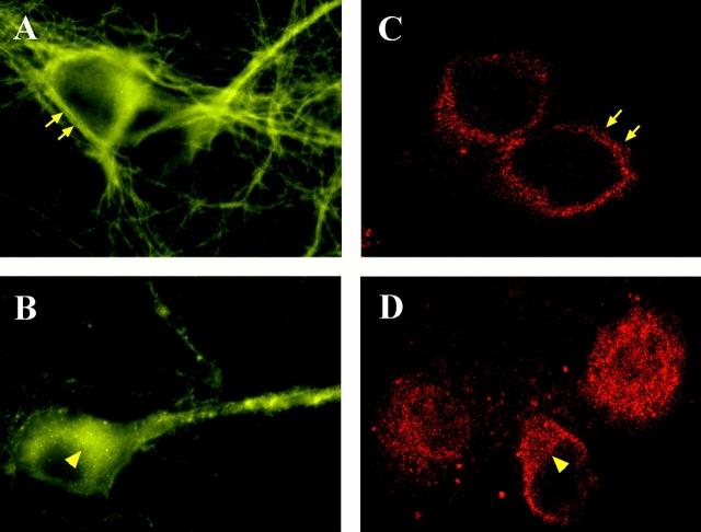
Figure 5.
Immunocytochemistry of hippocampal neurons stained by an antibody against the intracellular domain of TrkB under permeabilizing conditions. TBS was applied to the hippocampal neurons in the presence (B and D) or absence (A and C) of Cd2+ and kyn for 30 min. The cultures were then fixed with 4% paraformaldehyde for 30 min, permeabilized with 0.4% Triton X-100 for 60 min, and processed for immunofluorescence staining of TrkB. A and B are conventional immunofluorescence images, and C and D are confocal immunofluorescence images. Arrows indicate cell surface and arrowheads indicate cytoplasmic stainings, respectively. Note that active cells (stimulated by TBS) exhibit TrkB receptors mostly on the cell surface, whereas inactive cells (TBS plus Cd2+ and kyn) show more cytoplasmic staining of TrkB.

