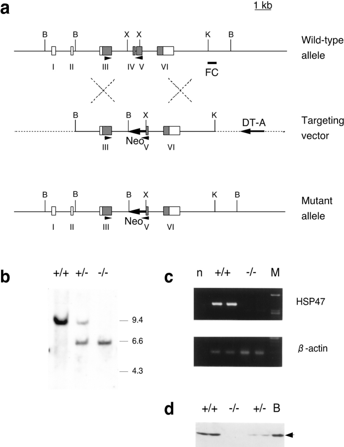
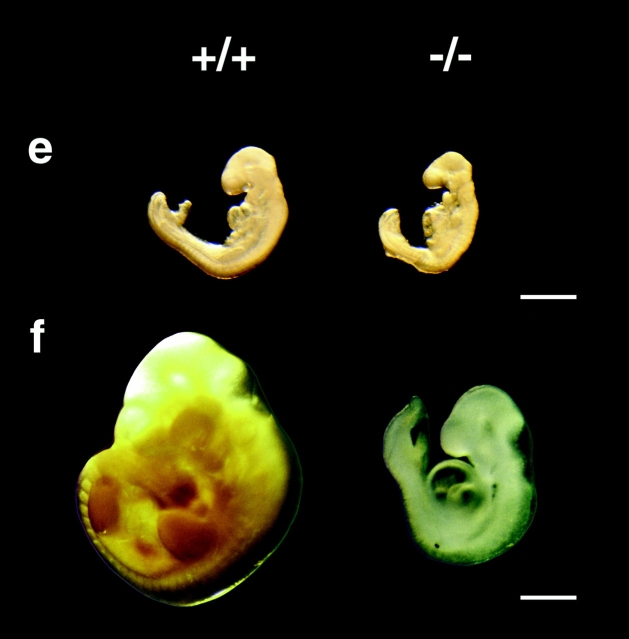
Figure 1.
Targeted disruption of the Hsp47 gene and characterization of the null phenotype. a, Homologous recombination with the targeting vector deletes exon IV and part of exon V, and simultaneously inserts a neomycin-resistance gene. Arrows indicate the orientation of neomycin-resistance gene and DT-A cassettes. Arrowheads indicate the location of primers used in RT-PCR assay. B, BamHI; K, KpnI; X, XhoI. b, Southern blot analysis demonstrating the genotypes of the offspring. A 3′ flanking probe shown as FC in a detects a 9-kb BamHI fragment in wild-type genomic DNA and a 6-kb fragment in the targeted allele in mice. Targeted ES clones were confirmed using a 5′ external probe (not shown). c, RT-PCR analysis was performed for the offspring using primers shown in a. n, Denotes the lane of negative control in which reactions are performed without RNA. d, Immunoblot analysis of Hsp47 was performed for wild-type, heterozygotic, and homozygotic null mice. B shows the positive control lane with the extract of Balbc/3T3 cells. Lateral views of 9.5 dpc (e) and 10.5 dpc (f) Hsp47−/− homozygous embryos (right) and wild type (left). Embryos were observed before (f) and after (e) fixation with 10% formaldehyde. Bars, 1 mm.


