
Figure 3.

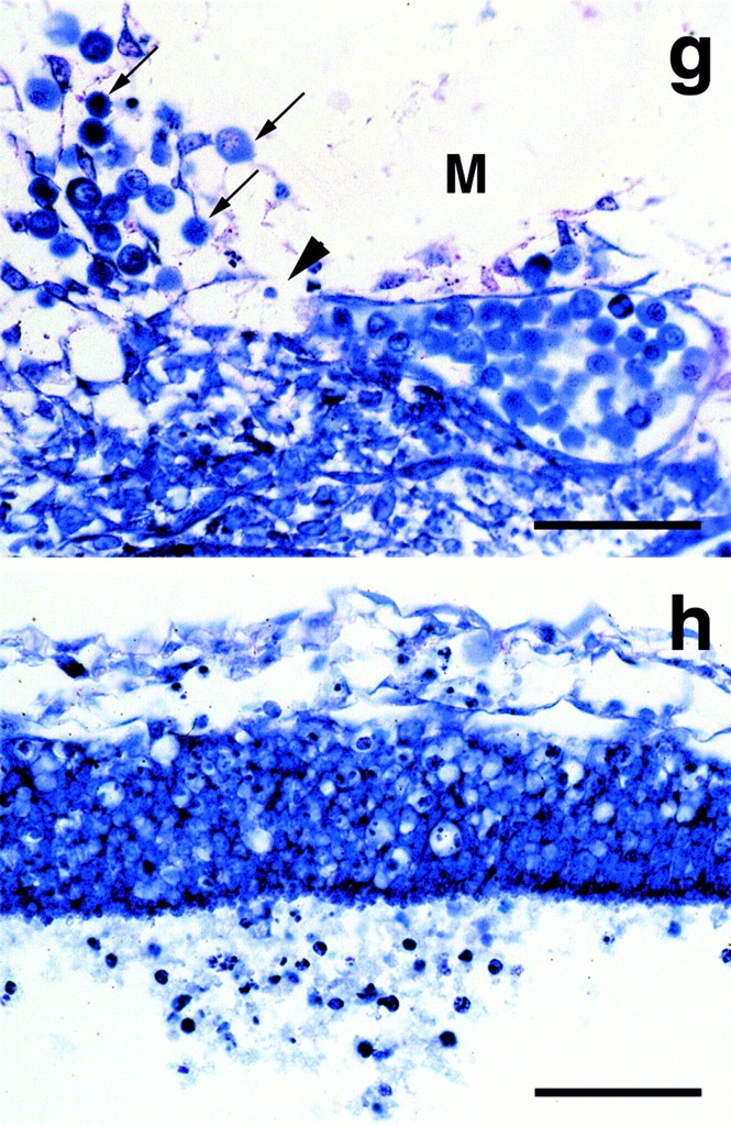
Silver impregnation analysis of 9.5 dpc wild-type (a and c) and homozygous (b and d) embryos. Neuroepithelium and underlying mesenchyme (a and b), and surface ectoderm and underlying mesenchyme (c and d) were analyzed. Hsp47−/− embryos had poorly developed reticular fibers in their mesenchymal tissues (b and d) compared with wild-type embryos (a and c). The basement membrane was hardly detected between the neuroepithelium of the neural tube and the surrounding mesenchymal tissue (b) of the Hsp47−/− embryos, whereas it was clearly detected as black staining in wild-type embryos (a). Neuroepithelium organization was disordered and cell division (arrowheads) was abnormally (closer to the mesenchymal tissue) detected in the cell layer of the neuroepithelium in Hsp47−/− embryos (b). Bars, 20 μm. Electron microscopic observation of the ectoderm in wild-type (e) and Hsp47−/− embryos (f). In e, a bundle of collagen fibrils (arrow) can be seen to reach out towards the lamina densa and to be distributed in the lamina fibroreticularis. The lamina densa (arrowhead) appears as a continuous sheet with a width of ∼30 nm positioned under the ectoderm (E) and the lamina lucida. The lamina densa (arrowhead) of the ectoderm (E) was interrupted (double arrowhead) and collagen fibrils were hardly discernible in Hsp47−/− embryos (f). The lamina densa seen in Hsp47−/− embryos was 30–50 nm in width, thicker than that in Hsp47+/+ embryos. Bar, 100 nm. Giemsa staining of Hsp47−/− embryos of 10.5 dpc shows the discontinuity of endothelial cells surrounding blood vessels (g, arrowhead) and the leakage of erythropoetic cells (g, arrows). Neuroepithelial cells also were also shown to leak out of the neural tube (h) by Giemsa staining. Bar, 50 μm.