
Figure 9.
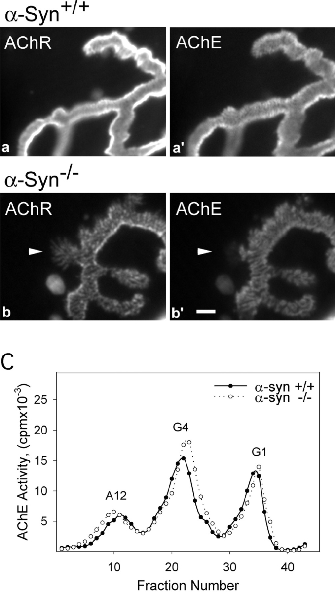
AChE in wild-type and α-Syn−/− NMJs. Sternomastoid NMJs from wild-type and α-Syn−/− mice were double-labeled for AChR and AChE, and imaged en face under identical microscope settings. The intensities of both labels are substantially reduced in the null NMJs (b and b′) compared with wild-type (a and a′). The AChE distribution in null NMJs shows most of the alterations found in the AChR distributions, except that AChE does not extend into the thin fingers of AChR (arrowhead in b and b′). Bar, 2 μm. C, The muscles from α-Syn−/− mice show no apparent differences in the synthesis and assembly of AChE. The AChE from sternomastoid muscles of α-Syn+/+ and α-Syn−/− mice was extracted and analyzed by velocity sedimentation. G1, Monomeric AChE; G4, tetrameric AChE; A12, synaptic form of AChE consisting of three tetramers attached to a collagen-like tail.