
Figure 1.
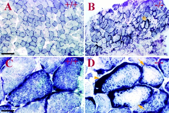
Alteration of mitochondrial distribution in desmin-null soleus skeletal muscle. SDH histochemical staining (A–D) demonstrates alteration of mitochondrial distribution in desmin-null (−/−) soleus muscle. In sections of normal (+/+) 10-mo-old mouse soleus muscle (A), a muscle consisting mostly of highly oxidative slow twitch fibers, a checkerboard pattern of intensely staining fibers is observed, with a variable amount of fibers staining with less intensity. The staining is mostly granular throughout the fibers, with slight enhancement of subsarcolemmal staining in a few fiber sections (C). In soleus sections from corresponding desmin-null animals (B, same scale as A), this discernible checkerboard pattern is partially lost. Large clumps of subsarcolemmal staining appear in a majority of the fibers (arrowhead), and the interior of fibers frequently show reduced SDH staining (arrows) (C and D, higher magnification view of A and B accordingly).