Figure 6.
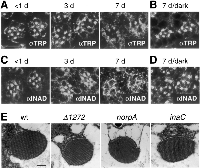
Spatial distribution of TRP and INAD in trpΔ1272 flies. Tangential sections of adult compound eyes shown in A–D were obtained from trpΔ1272 at the indicated ages (<1, 3, and 7 d after eclosion). (A) Sections were prepared from flies reared under a normal light/dark cycle and stained with anti-TRP antibodies (αTRP). (B) Sections were prepared from 7-d-old flies maintained constantly in the dark and stained with anti-TRP antibodies (αTRP). (C) Sections were prepared from flies reared under a light/dark cycle and stained with anti-INAD antibodies (αINAD). (D) Sections were obtained from 7-d-old dark-reared flies and stained with anti-INAD antibodies (αINAD). (E) Ultrastructure of single rhabdomeres from 1-d-old wild-type (wt), trpΔ1272, (Δ1272), norpA, and inaC flies viewed by transmission electron microscopy. Bars: (A) 10 μm; (E) 0.5 μm.

