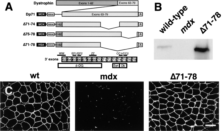
Figure 1.
Generation of dystrophin Δ71–78 transgenic mdx mouse. (A) Shown are the dystrophin transgenic expression vectors that contain Dp71 and full-length cDNAs deleted for the indicated exons. The binding sites for β-dystroglycan (β-DG), syntrophin (Syn), and dystrobrevin (Db) are delineated relative to the exon boundaries of the dystrophin gene. Exon 79 encodes only three amino acids and therefore is not shown. Dystrophin structural motifs are also indicated that include the WW, EF1, EF2, ZZ, and coiled coil (CC) 1 and 2 domains. The first half of the WW domain is encoded on exon 62. All constructs contain either a mouse muscle creatine kinase [MCK] or the HSA promoter, the SV40 VP1 intron (intron), and the SV40 poly-adenylation site (“A”). (B) Western blot analysis of total skeletal muscle proteins from wild-type, mdx, and dystrophin Δ71–78 mice using an NH2-terminal specific dystrophin antibody. (C) Immunofluorescent staining for dystrophin in muscle quadriceps reveals uniform expression in wild-type and dystrophin Δ71–78 mice. Scale bar, 50 μm.

