
Figure 4.
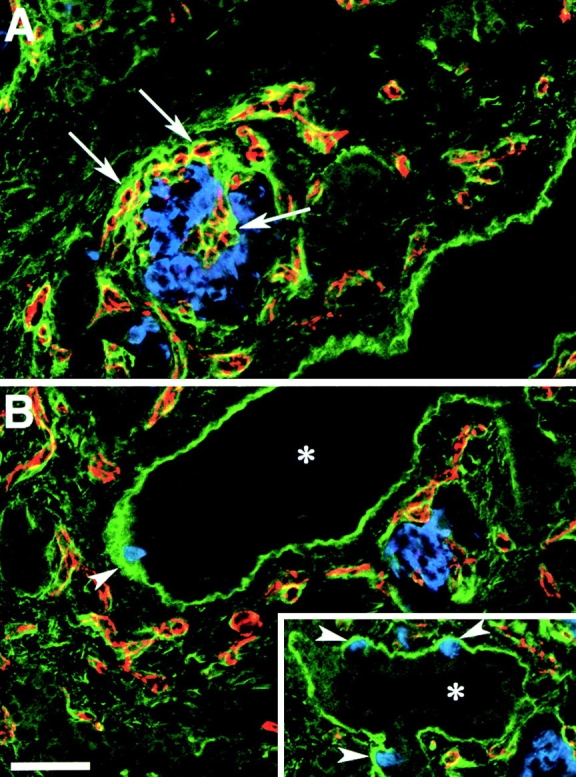
FN expression pattern in the developing human pancreas. Confocal images of two adjacent microscopic fields (A and B) from cryostat sections stained for FN (green), insulin (blue), and PECAM-1 (red). A strong FN-specific immune reactivity (green) highlights the basal membrane of ducts (asterisk) and of insulin-positive cells (blue) emerging from the ductal epithelium (arrowheads, B and inset). The PECAM-1-specific staining (red), identifying blood vessels, reveals that the basal membrane of endothelial cells, infiltrating developing islet clusters, is also marked by a bright FN-specific staining (arrows). Bar, 100 μm.