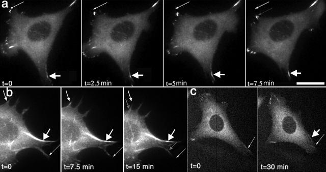
Figure 10.
Clusters of paxillin and α-actinin translocate along the lateral edge of the cell in areas of cell retraction. (a) In retracting regions of the cell, paxillin-GFP clusters move centripetally along the edge of the cell (thin arrows). Strong lateral clusters (thick arrow) strengthen as smaller adhesive structures incorporate into them but move slower than the smaller clusters (thin arrows). (b) α-Actinin–GFP also resides in clusters along the cell edge that move centripetally (thin arrows). The smaller clusters move faster than the larger clusters (thick arrow). (c) Paxillin-GFP clusters remain intact for over 30 min as they translocate along the cell edge. Compare the original location of the cluster marked with a thin arrow to the new location marked with a thick arrow. Videos 8–10 available at http://www.jcb.org/cgi/content/full/153/7/1427/DC1. Bar: (a) 5 μm; (b) 4.5 μm; (c) 8.7 μm.

