
Figure 1.
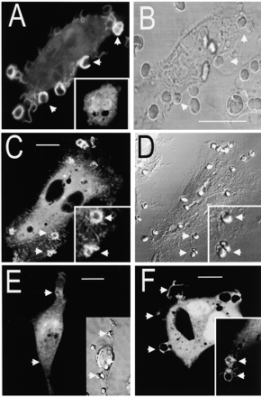
Distribution of PH domain–GFP chimeras in macrophages. RAW 264.7 cells were transfected with chimeras of GFP and the indicated PH domain and then exposed to IgG-opsonized SRBCs to initiate phagocytosis. Confocal fluorescence (A, C, E, and F) and DIC images (B, D, and E, inset) were acquired. (A–B) Cells transfected with Akt-PH–GFP. The inset shows the distribution of Akt-PH–GFP before addition of SRBCs. (C–D) Cells transfected with Gab1-PH–GFP. (E) Cells transfected with Akt-PH–GFP were pretreated with 100 nM wortmannin for 15 min before addition of the SRBCs. (F) The main panel shows cells transfected with (R28C)Btk-PH–GFP, a mutant form of Btk-PH–GFP unable to bind 3′PI in vitro. The inset shows a cell transfected with wild-type Btk-PH–GFP. Arrows indicate sites of attachment of SRBCs. Images are representative of at least three experiments of each type. Bars, 10 μm.