
Figure 8.
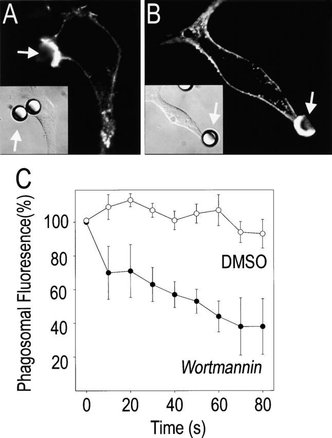
Reversal of 3′PI accumulation by wortmannin. (A and B) RAW 264.7 cells were allowed to interact with IgG-opsonized polystyrene beads (8-μm diameter), and when phagocytosis commenced the cells were treated with either 100 nM wortmannin or with an equivalent volume of the vehicle (DMSO). After 60 s, the cells were fixed, and F-actin was stained with rhodamine-phalloidin. A and B show F-actin distribution, and insets show the corresponding DIC image. (A) DMSO-treated; (B) wortmannin-treated. In C, the cells were transfected with Akt-PH–GFP ∼24 h before exposure to the beads. The concentration of the chimera at the phagosomal cup was monitored by fluorescence microscopy, and when a sizable accumulation occurred the samples were treated with wortmannin or DMSO. The persistence of Akt-PH–GFP was monitored thereafter at 10 s intervals. Data were normalized to the fluorescence at the time of addition of the inhibitor to facilitate comparison. Data are mean ± standard error of three experiments.