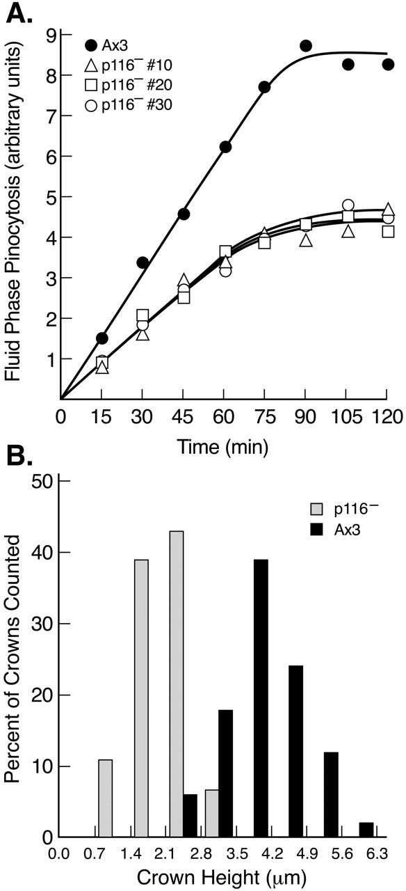
Figure 9.

Rate of fluid phase endocytosis, and crown size, in wild-type and p116− cells. (A) Kinetics of accumulation of the fluid phase pinocytic marker FITC-dextran (arbitrary units) within three independent p116− cell lines and the parental line Ax3 (see key). Each value is the mean of duplicate samples. (B) Histogram of the height of crowns in Ax3 and p116− cells (see key). The values, which were taken from confocal Z series of cells stained for actin, were binned in 0.7-μm intervals and are presented as a percent of total crowns counted (64 in Ax3, 58 in p116−). Crowns were counted only if they rose almost vertically from the dorsal surface of cells. The bottom of such crowns typically exhibited a spherical, solid disc of actin staining. Above this the crown had the appearance of a ring of fluorescence in each section. Similar results were obtained using cells stained for coronin.
