
Figure 5.
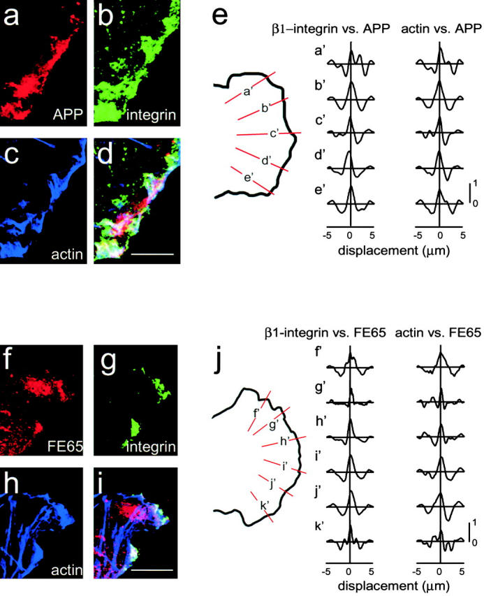
APP and FE65 colocalize with β1-integrins and actin in lamellipodia. (a–e) H4 cells triple labeled with APP polyclonal antibodies (a), β1-integrin monoclonal antibody (b), and Oregon green–conjugated phalloidin (c). Overlap of all three proteins is indicated by white in the overlay (d). (f–j) H4 cells triple labeled with FE65 polyclonal antibody (f), β1-integrin monoclonal antibody (g), and Oregon green–conjugated phalloidin (h). Overlap of all three proteins is indicated by white in the overlay (i). (e and j) Cross-correlation analysis of APP and FE65 colocalization with β1-integrins and actin in focal complexes. Lines were drawn perpendicular to the lamellipodial edge (shown in orange and labeled a′–e′ for APP and f′–k′ for FE65). The intensities were determined along each line and the cross-covariograms calculated as described in Materials and Methods. All of the cross-covariograms have peaks >0.5, indicating significant correlation. None have a displacement greater than the half-width at half-height, indicating colocalization. Bars, 10 μm.