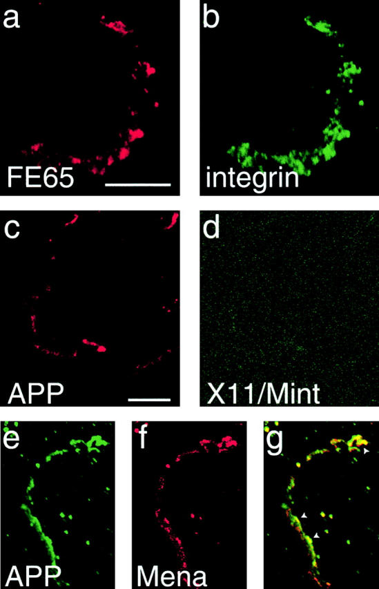
Figure 6.

FE65 and APP remain associated with focal complexes left behind after detachment of lamellipodia. (a and b) H4 cells were labeled with polyclonal FE65 antibodies and monoclonal β1-integrin antibody. Where a lamellipodium was detached from the substrate, the integrin-dependent focal complex was left behind and labeled with both FE65 antibodies (a) and integrin antibody (b). (c and d) H4 cells were labeled with APP polyclonal antibody and X11/Mint monoclonal antibody. Where lamellipodia were detached from the substrate, adhesion sites left behind were labeled with APP antibody (c), but not with X11/Mint antibody (d). (e–g) Similar adhesion sites that are double labeled with APP monoclonal antibodies (e) and Mena polyclonal antibody (f). Overlap between APP and Mena in adhesion sites is indicated by yellow in the overlay (g). Arrowheads point to areas that contain high levels of both APP and Mena. Bars, 10 μm.
