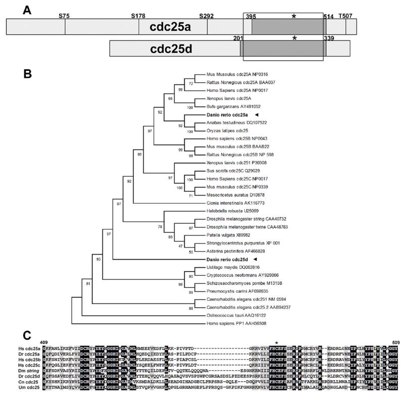
Figure 1.
a: Schematic of cdc25a and cdc25d from zebrafish, to scale. Conserved Chk1 phosphorylation target sites from human Cdc25A are shown as vertical lines, labeled according to the residue in human Cdc25A. Boxed region shows alignment in figure 1c and dark box delineates cdc25 phosphatase domain as defined by NCBI conserved domain search. Asterisk indicates position of HCXXXXXR catalytic motif. b: UPGMA phylogenetic reconstruction of cdc25 genes from various species. Accession numbers used are noted in the figure. c: Alignment of the phosphatase domain of Cdc25 proteins: Dr Danio rerio Dm Drosophila melanogaster Hs Homo sapiens Cn Cryptococcus neoformans Um Ustilago maydis. Accession numbers are as in panel a.

