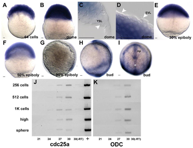
Figure 4.

In situ hybridization using a cdc25a antisense probe during epiboly (a–i). Panels represent staged semi-quantiative RT-PCR results using either cdc25a (j) or ODC (k) specific primers. Cycle numbers are as indicated below panels, with stages at left. (+) plasmid positive control; (-RT) no RT control reactions. All embryos are oriented animal pole up. g and h are lateral views with the dorsal side to the right and i is a dorsal view. YSL is yolk syncitial layer, EVL is enveloping layer, n is notochord and np is neural plate. Scale bar is 20μm.
