
Figure 3.
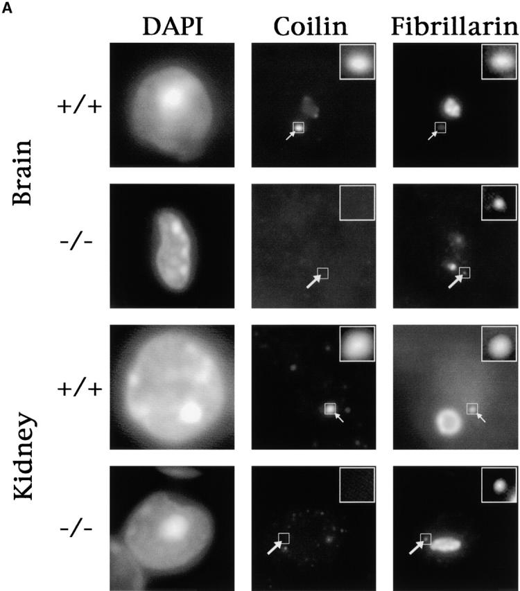

Knockout mice display “residual” CBs. (A) Frozen tissues were sectioned and stained with antibodies against coilin and Fb. In wild-type tissues, coilin is colocalized with Fb in CBs (small arrows) and sometimes is visible in nucleolar caps (see wild-type brain panels). Coilin staining is absent from knockout tissues, but residual CBs (large arrows) are evident as extranucleolar Fb foci. For better visualization, insets display the boxed CBs or residual CBs at higher magnification. (B) Confocal imaging of sensory neurons from dorsal root ganglion of a knockout animal costained for coilin/Gemin2, Nopp140/SMN, or Nopp140/U2B′′. Note the residual CBs denoted by white arrows in the Nopp140 channel and the absence of SMN or U2B′′ within these structures. (C) Sensory ganglion neurons from wild-type and knockout animals were stained with silver to visualize nucleoli and CBs (black arrows). Note the absence of extranucleolar silver deposition in the knockout cells.