
Figure 5.
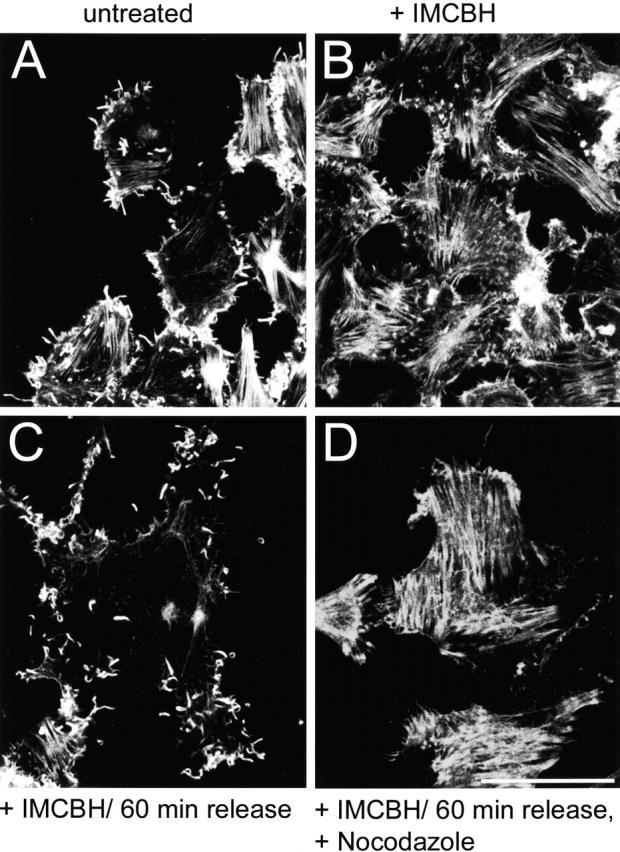
Microtubules and IEV particles are required for the formation of actin tails. RK13 cells were infected with VV at 10 PFU/cell in either the absence (A) or presence (B–D) of IMCBH. At 8 hpi, the cells in C and D were washed three times in DME (C) or in DME containing 33 μM nocodazole (D). 1 h later, all cells were fixed and stained with rhodamine-conjugated phalloidin as described in Materials and methods. Samples were analyzed by confocal microscopy, and the reconstructed z-series are shown. Bar, 10 μm.