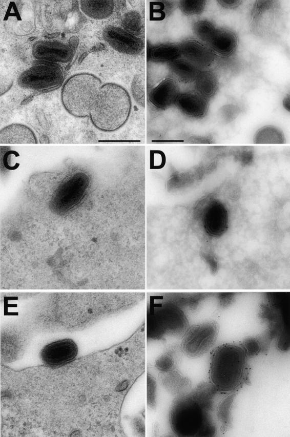
Figure 8.

VV morphogenesis in cells infected with vB5R-GFP. RK13 cells were infected with vB5R-EGFP and processed for conventional EM after fixation at 12 hpi (A, C, and E). Alternatively, samples were processed for cryo-immunoelectron microscopy and labeled with anti-GFP and protein A gold (B, D, and F). Bars, 300 nm.
