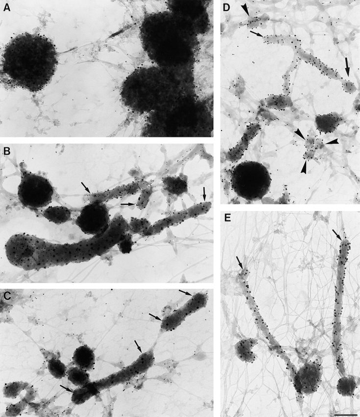
Figure 3.

MIICs in immature and maturing D1 cells. Cells were loaded with HRP for 1 h in the absence of LPS as in the legend to Fig. 2 A (A) or 1 h in the presence of HRP plus 6 h HRP + LPS (B and C) or pulsed for 1 h with HRP and chased for 3 h in the presence of LPS (D and E) and processed for whole-mount EM. (A) Vacuolar MIICs in immature D1 cells labeled for MHC II (10 nm gold) and DM (15 nm gold). Tubular MIICs have formed after 3 (D and E) or 6 h (A and B) of LPS treatment. (B) Double labeling for MHC II (10 nm gold) and DM (15 nm gold). A tubule seems to form out of the vacuole in the left bottom corner. (C) Tubular MIICs labeled for MHC II (10 nm gold) and LAMP-1 (15 nm gold), demonstrating the late endosomal/lysosomal character of these organelles. (D) Tubular MIICs and free vesicles (arrowheads) with diameters ranging from 80 to 200 nm were double labeled for MHC II (10 nm gold) and DM (15 nm gold). Note the vesicle that seems to be formed at the tip of a tubule (thick arrow). (E) Tubular MIICs, showing DM (10 nm gold) and LAMP-1 (15 nm gold). Note the cytoskeletal elements in which the MIICs are embedded.
