Figure 3.
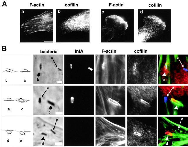
Cofilin is recruited to InlB-induced membrane ruffles and phagocytic cups. (A) Colocalization of cofilin with F-actin in InlB-induced ruffles. Vero cells, untreated (a and b) or stimulated with 4.5 nM of InlB (c and d) stained with FITC-phalloidin and anti-cofilin Ab. (B) Recruitment of cofilin by bacteria at different stages of the internalization process in Ref52 cells. The left panels schematically represent the bacteria detected in the five other panels and marked by reference to Fig. 1 A. Total bacteria associated with cells was visualized in phase–contrast, and bacteria that are fully or partly extracellular were identified by labeling with anti-InlA Ab before cell permeabilization. Cells were stained with FITC-phalloidin and anti-cofilin Ab. In the merged images, extracellular bacteria are blue, F-actin is green, and cofilin is red. Arrowheads or arrows indicate bacteria that colocalizes or not with cofilin, respectively. Bars: (A, a) 10 μm; (A, c) 2 μm.

