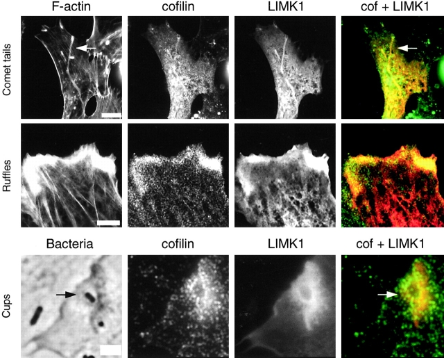
Figure 7.
In LIMK1-overexpressing cells, endogenous cofilin is still recruited to actin-based structures. Vero cells transiently transfected with LIMK1 were analyzed for Listeria tail formation, InlB-induced ruffling, or bacterial entry as described in the legends to Figs. 4 and 5. Cells were stained with FITC-phalloidin, anti-Myc, and anti- cofilin Ab. In the merged images, cofilin is green and LIMK1 is red. Bars: (first and second rows) 10 μm; (third row) 2 μm.

