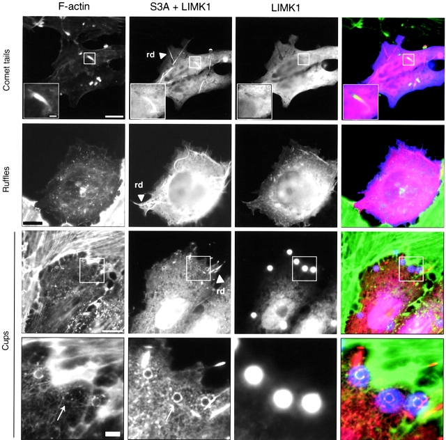
Figure 8.
Overexpression of S3A cofilin suppresses F-actin accumulation induced by LIMK1 overexpression. Vero cells transiently cotransfected with LIMK1 and S3A cofilin cDNAs were analyzed for formation of Listeria tails, InlB-induced ruffles, or InlB bead phagocytic cups as described in the legends to Figs. 4 and 5. Cells were stained with FITC-phalloidin, anti-Myc, which detects both LIMK1 and S3A fusion proteins and lights up cofilin rods (arrowheads, rd), and an antipeptide Ab specific only to the LIMK1 fusion protein. For cups, boxed regions indicate the position of the field, which is magnified below. Due to their intrinsic fluorescence in the 650–700-nM range, InlB beads are detected together with the Cy5-labeled LIMK1 anti-tag. In the merged images, F-actin is green, Myc is red, and LIMK1 and InlB beads are blue. Bars: (full images) 10 μm; (magnified images) 2 μm.

