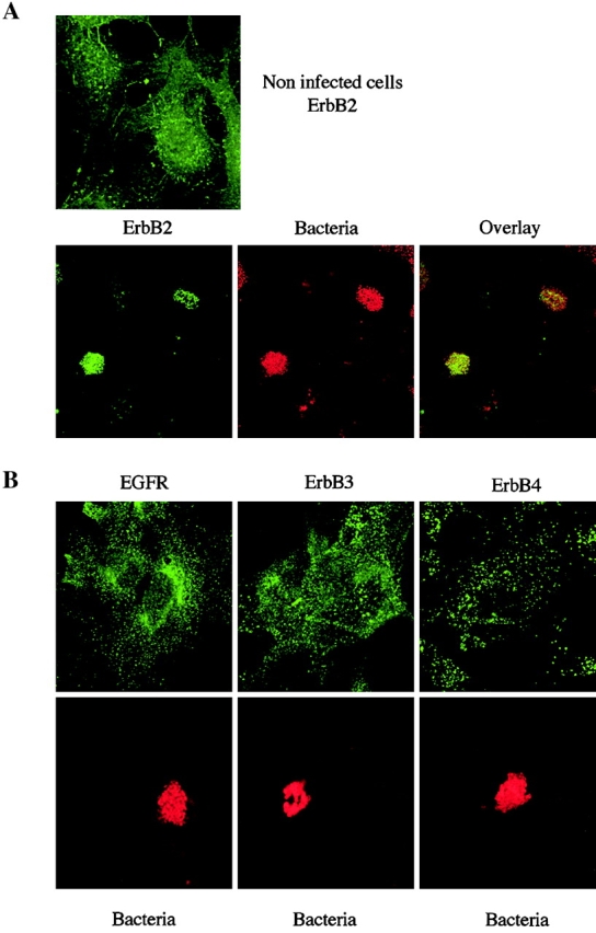
Figure 3.

Localized adhesion of N. meningitidis induces the clustering of ErbB2, but not of the other ErbB members. (A) Top, uninfected cells were stained for ErbB2; bottom, cells infected for 3 h with the 2C43 wild-type strain of N. meningitidis were double-stained for ErbB2 (left) and bacterial colonies (middle). Merged images (overlay) of the same fields are presented on the right. (B) Cells infected for 3 h with the 2C43 wild-type strain of N. meningitidis were double-stained for either EGFR, ErbB3, or ErbB4 (top, as indicated) and bacterial colonies (bottom). Panels show receptor and bacterial staining in the same fields.
