
Figure 1.
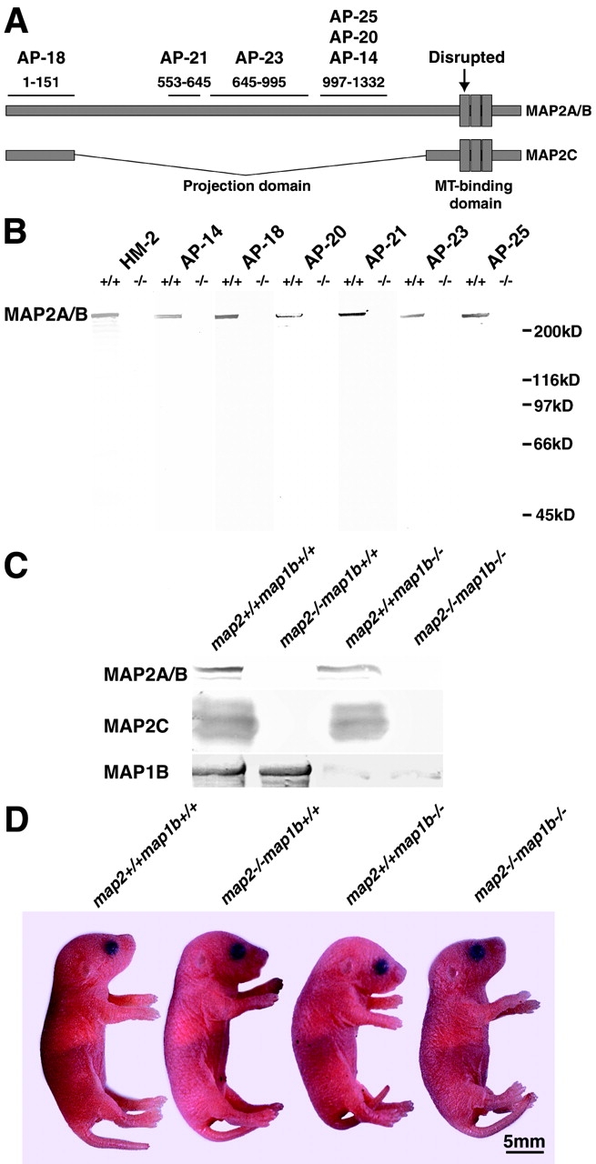
Targeted disruption of the map2 gene. (A) Diagrammatic representation of MAP2 structure. Epitopes of six anti-MAP2 mAbs (Kalcheva et al., 1994) are shown. The neo cassette (Harada et al., 1994) is inserted at the point indicated by an arrow. (B) Western blotting of crude extracts of the whole brain with a series of anti-MAP2 mAbs (HM2, AP-14, AP-18, AP-21, AP-22, AP-24, and AP-25). Mice at 8 w old were analyzed. Note the absence of MAP2 immunoreactivity in all map2 −/− lanes. (C) Immunoblot analysis of crude extracts from P0 mouse brain with the anti-MAP2 mAb HM2 and the anti-MAP1B antiserum 3d2. (D) Appearance of map2 +/+ map1b +/+, map2 −/−map1b +/+, map2 +/+ map1b −/−, and map2 −/−map1b −/− mice at P0.