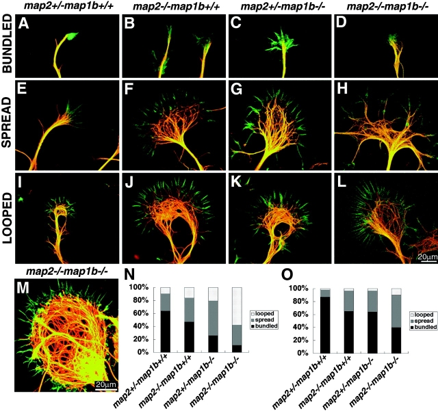
Figure 8.
Morphology of growth cones in cultured hippocampal pyramidal neurons. (A–M) Cells were double labeled with phalloidin (green) and a monoclonal antibody against tyrosine tubulin TUB-1A2 (red). Examples of bundled (A–D), spread (E-H), and looped (I-L) MTs in the growth cones after 2 d of culture of neurons from each genotype are shown. (M) Example of the abnormal cytoskeletal mass of growth cones from 8-d-old cultured neurons of map2 −/−map1b −/− mice. (N and O) Histograms of the percentages of three types of MT organization of the growth cones in axons (N) and minor processes (O).

