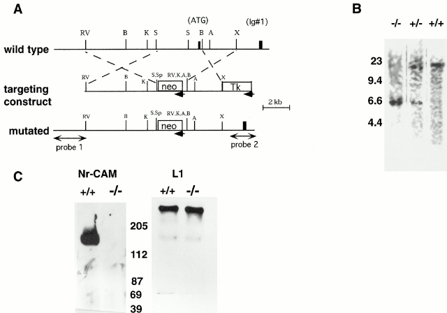
Figure 2.
Generation of Nr-CAM null mice. (A) Genomic organization of Nr-CAM gene and gene targeting construct. One clone containing the region from the first intron to forth exon was obtained by genomic screening. The second exon, which encodes the initiation codon (ATG) and signal sequence for Nr-CAM (black box, middle), was replaced with a neo cassette. Direction of transcription for neo is opposite to that of Nr-CAM as indicated by arrow. The black box at the right end represents the fourth exon, which encodes half of first Ig domain (Ig #1). Probes 1 and 2 were used for screening of targeted embryonic stem cells. (B) Southern blot analysis of genomic DNAs from 4-wk-old progeny. DNA was extracted from tail and digested with EcoRV. Nonradioisotope Southern hybridization with probe 2 was performed as described in Materials and methods. Wild-type allele gives ∼15-kbp band, whereas mutated allele gives ∼6-kbp band. DNA standards in kbp are shown on the left side. (C) Western blot analysis of adult brain extracts. 20 μg of extract were subjected to 7% SDS PAGE and blotted with anti–Nr-CAM and anti-L1 antibodies. Anti–Nr-CAM antibody recognizes a 140-kD band which corresponds to cleaved Nr-CAM extracellular region. No bands were detected in the −/− extract. Anti-L1 antibody recognizes a 200-kD band and weak 140-kD band both in wild-type and −/− extracts. Molecular mass markers in kD are shown at their respective positions between the two panels.

