
Figure 3.
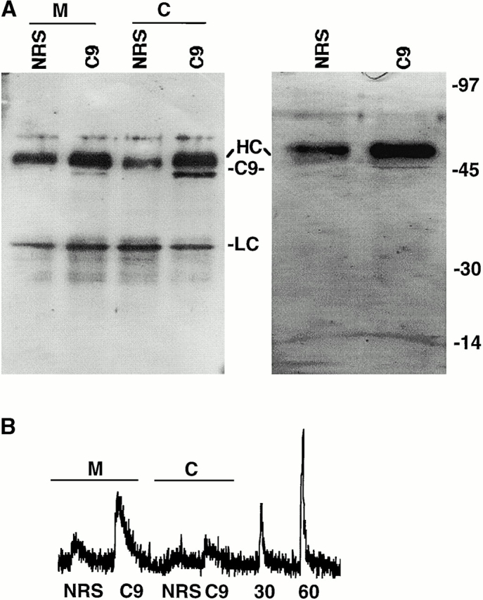
S-Nitrosylation of cytoplasmic and mitochondrial caspase-9. Caspase-9 immunoprecipitation. Proteins were immunoprecipitated from mitochondrial (M) or cytoplasmic (C) cellular fractions using a caspase-9–specific rabbit polyclonal antiserum (C9) or equal concentration of normal rabbit serum (NRS). Immunoprecipitated proteins were visualized on silver-stained gels (right) or caspase-9 Western blot analysis (left). In the silver-stained gel, immunoprecipitates from whole cell lysates are shown. Molecular weight markers, immunoglobulin heavy chain (HC), light chain (LC), and caspase-9 (C9) are shown. (B) S-Nitrosylation of caspase-9. The SNO-derived chemiluminescence signal of control normal rabbit serum (NRS) or caspase-9 (C9) immunoprecipitates obtained from mitochondrial (M) and cytoplasmic (C) fractions of BJAB cells and from 30 nM (30) and 60 nM (60) S-nitrosoglutathione standards are shown. The data are representative of 1 of 10 separate experiments.