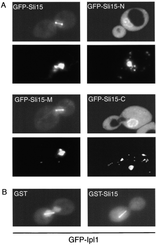
Figure 5.

Subcellular localization of GFP–Sli15 and GFP–Ipl1. (A) Subcellular localization of full-length and truncated forms of Sli15. Wild-type diploid yeast cells (DBY1830) expressing GFP–Sli15 (pCC1060), GFP–Sli15-N (pCC1533), GFP–Sli15-M (pCC1534), or GFP–Sli15-C (pCC1566) were stained with DAPI and examined. For each pair of images: top, GFP fusion protein image; bottom, DAPI-stained image. (B) Subcellular localization of GFP–Ipl1. Wild-type haploid yeast cells (TD4) carrying pCC1584 (GFP–Ipl1) and pEG(KT) (GST) or carrying pCC1584 and pCC1061 (GST–Sli15) were cultured for 2 h in medium containing 4% galactose to induce expression of GST or GST–Sli15, respectively. Images of GFP–Ipl1 are shown.
