Figure 5.
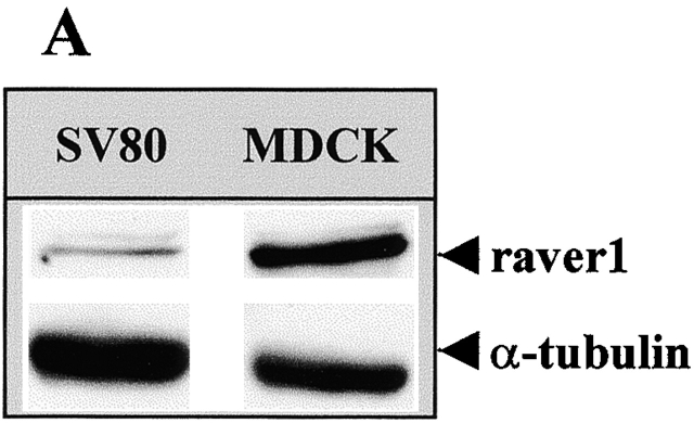
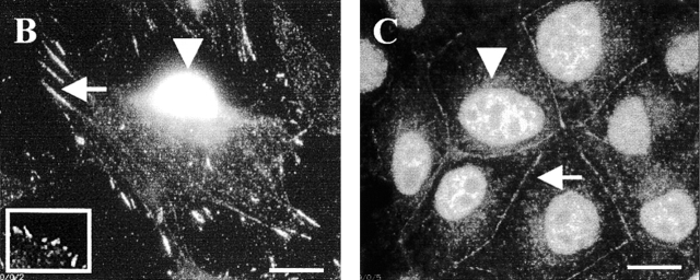
Raver1 is a dual compartment protein. (A) Western blot analysis of raver1 and α-tubulin. Normalized protein samples of MDCK and SV80 cells were resolved by SDS-PAGE and immunoblotted with either raver1 or α-tubulin antibodies. Note that MDCK epithelial cells express raver1 to an ∼10-fold higher level than SV80 fibroblasts. (B and C) Immunofluorescence analysis of SV80 fibroblasts (B) and MDCK epithelial cells (C). Cells were fixed and subsequently stained with the monoclonal raver1 antibody. Note the strong nuclear signal for raver1 (B and C, arrowheads) in addition to the location at large, probably mature (B, arrow) and small peripheral focal contacts (B, inset) of the fibroblasts and at cell–cell contacts of epithelial MDCK cells (C, arrow). The exposure time of the picture in B was optimized for revealing focal contact staining, resulting in an overexposed nuclear image in the SV80 cell. Bars, 10 μm.


