
Figure 5.
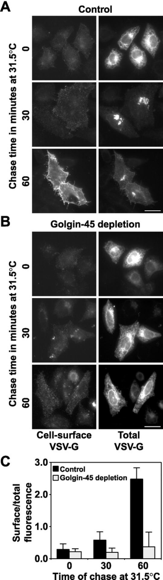
Depletion of golgin-45 disrupts transport of VSV-G protein from the ER to the cell surface. Control (A) or golgin-45 (B) RNAi cells were transfected with a plasmid encoding GFP-tagged VSV-G ts045 protein. VSV-G ts045was arrested in the ER at 39.5°C and then chased out at 31.5°C for 30 and 60 min. Images are shown of cells fixed after 0, 30, and 60 min of chase for both total and cell surface–associated VSV-G. (C) The extent of VSV-G transport after 0, 30, and 60 min chase at 31.5°C was measured in control and golgin-45–depleted cells. This ratio does not approach unity due to the different dyes used to measure surface and total fluorescence. The data shown is representative of three experiments with n = 20 for all data points in each experiment. Live cell videos showing VSV-G ts045 transport in control and golgin-45 depleted cells are available at http://www.jcb.org/cgi/content/full/jcb.200108079/DC1. Bars, 20 μm.