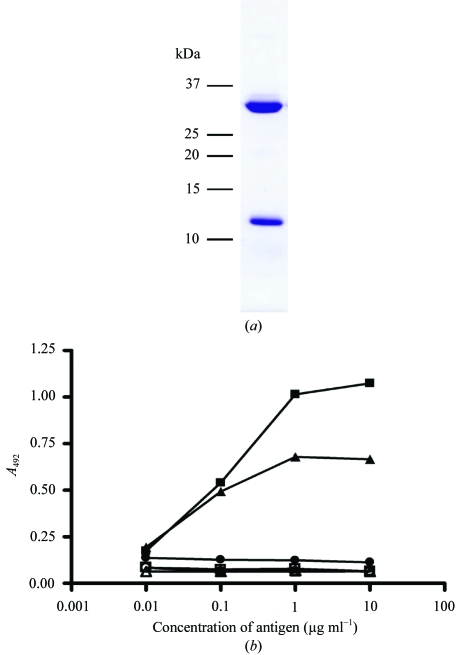
Figure 1.
HLA-G refolding and functional testing. (a) SDS–PAGE analysis of refolded HLA-G–RIIPRHLQL complex following anion-exchange and size-exclusion chromatography. (b) ELISA of refolded HLA-G. Two antibodies specific for human MHC molecules were used in a sandwich ELISA with HLA-G (filled squares) and HLA-B8 (filled triangles) as positive controls and LC13 (filled circles) as a negative control. Open symbols indicate the control experiment without primary antibody. Data represents the mean of three experiments.

