Figure 1.
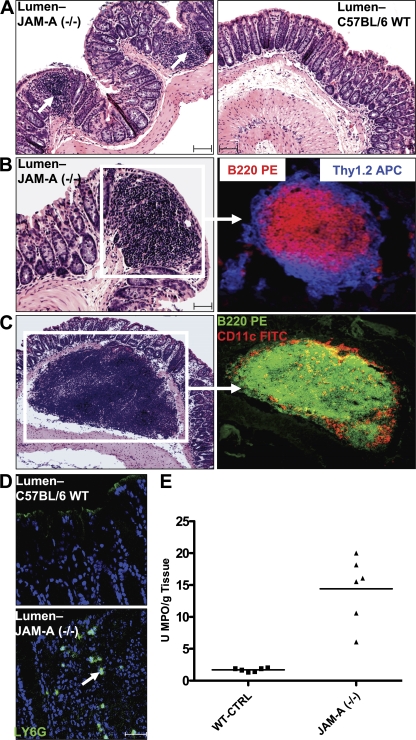
Increased colonic inflammation in JAM-A−/− mice. (A) Increased small lymphoid aggregates in JAM-A−/− mice. (B) Large lymphoid aggregates in JAM-A−/− mice identified as isolated lymphoid follicles. (C) Large, isolated submucosal lymphoid follicles observed only in JAM-A−/− mice. CD11c staining is shown in red. (D) Increased PMN in the colonic mucosa of JAM-A−/− mice. Bars, 40 μm. (E) MPO activity in the colonic mucosa of JAM-A−/− mice and littermate controls (n = 6). Horizontal bars represent the mean.

