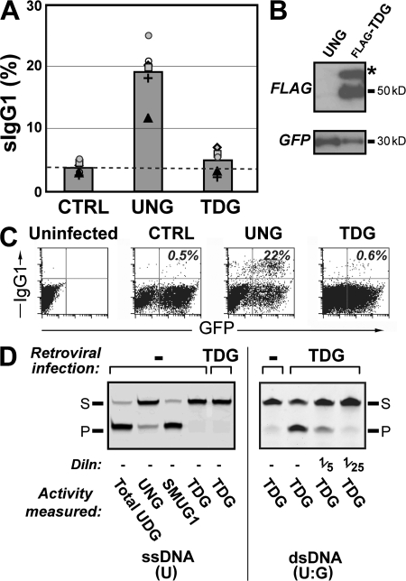
Figure 6.
Retroviral expression of TDG does not restore class switching in ung−/− B cells. (A) Comparison of switching to IgG1 by B cells from ung−/− mice that have been infected with the pMX retroviral vector encoding GFP alone or GFP together with either UNG or FLAG-tagged TDG, as monitored in multiple experiments indicated by the different symbols. The difference between the switching obtained with TDG and that obtained in the CTRL sample is not significant. (B) Analysis of FLAG-TDG expression in retrovirally infected B cells as monitored by Western blot using an anti-FLAG mAb; the band marked by an asterisk is probably sumoylated TDG (37). A Western blot for the vector-encoded GFP provides a control for infection efficiency. (C) Flow cytometric plots showing switching to IgG1 in purified B cells from an ung−/− msh2−/− mouse that has been infected with the pMX retroviral vector encoding GFP alone or GFP together with either UNG or FLAG-tagged TDG. Switching was assayed as in Fig. 1, with the proportion of retrovirally infected (GFP+) cells that have switched to IgG1 indicated in the top right quadrants. (D) Demonstration of TDG-encoded uracil-excision activity in extracts of retrovirus producing cells that have been transfected with the pMX retroviral vector encoding FLAG-tagged TDG. (left) Uracil-excision activity was monitored on a U-containing single-stranded oligonucleotide (which is not a substrate for TDG), whereas a double-stranded oligonucleotide containing a U:G mismatch is used on the right. Because TDG, UNG, and SMUG1 will all act as uracil-DNA glycosylases (UDG) on such a dsDNA oligonucleotide substrate, extracts were preincubated with Ugi (to inhibit UNG) and the PSM1 mAb (to inhibit SMUG1) when wishing to restrict the assay to TDG.

