Figure 1.
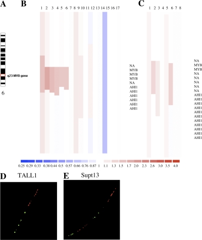
The MYB gene is tandemly duplicated in human T-ALL cell lines and patient samples. (A) Ideogram of chromosome 6 showing location of the MYB gene on q23. (B) Array CGH performed on DNAs from 17 T-ALL cell lines with Human Genome CGH 44K Microarrays (Agilent). A localized region on 6q23 surrounding the MYB locus is shown to have increased copy number in six of the cell lines. A 2.3-Mb region of chromosome 6 is shown. The top is centromeric, and the bottom is telomeric. Part of the AHI1 gene directly downstream of MYB is also amplified. NA indicates a probe in an intergenic region. Red indicates increased copy, blue indicates decreased copy, and the intensity of the color reflects the level of increase or decrease. (C) DNAs from leukemic cells in the diagnostic bone marrow of eight T-ALL patients were similarly analyzed by array CGH using Human Genome CGH 244K Microarrays, with an increased MYB copy number identified in two cases. A 500-kb region of chromosome 6 is shown. (D) Fiber-FISH on a T-ALL (TALL1) cell line with a diploid MYB copy number. The fosmid encompassing most of the MYB gene is labeled in green; a fosmid immediately 3′ of the MYB coding sequence is labeled in red. (E) Fiber-FISH on the Supt13 cell line showing a duplication of both fosmids spanning the entire MYB locus oriented in tandem on the same DNA fiber.

